MetIA — Deploying a Deep Learning Interface for Brain Metastases Segmentation
In radiotherapy treatment planning, clinicians manually contour the brain lesions to be treated on MRI — a slow, repetitive task. A deep learning model developed during an earlier Master’s project at IDM Caen offered a way to automate this delineation; my job was to take that model out of the lab and into clinical practice.
As a Research and Development Intern — Medical AI at the Centre François Baclesse (27/05/2024–05/07/2024), under the supervision of Dr. Corroyer-Dulmont (Head of AI and Medical Imaging Engineer at CFB), I built the visualization interface and API around the model and got it running on the center’s infrastructure.
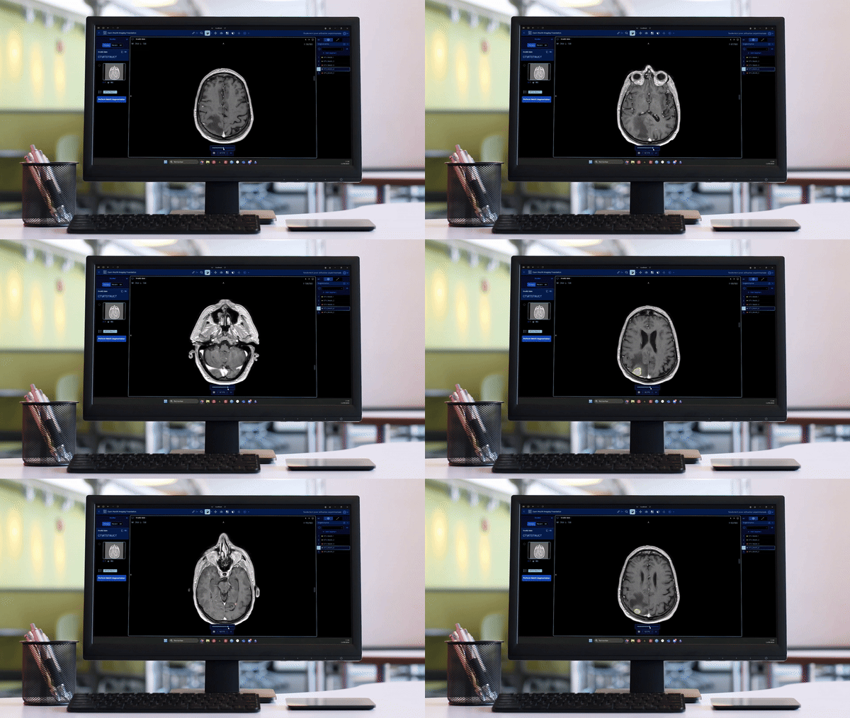
What I did
- Deployed, finalized, and optimized an interface based on the OHIF Viewer and its API on the center’s Windows environments — the ML Ops side of shipping a deep learning solution into a clinical workflow.
- Optimized the deep learning model to reduce its computational cost.
- Retrained the model on a new dataset.
- Built the API layer (Flask) connecting the OHIF front-end to the segmentation model.

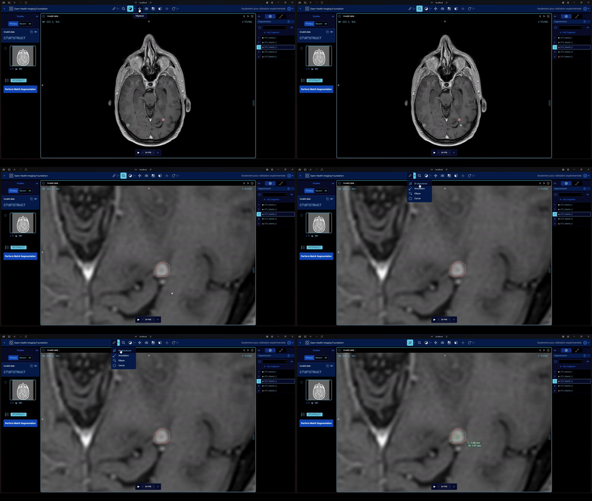
Skills strengthened
Working with DICOM and NIfTI-format MRI/CT data end-to-end, understanding a complex neural architecture (UNETR) well enough to retrain and optimize it, and adapting a research prototype to a clinical, multi-OS deployment environment.
Published outcome
This work fed directly into a peer-reviewed publication: I’m co-first author on “Development and routine implementation of a deep learning algorithm for automatic brain metastases segmentation on MRI for RANO-BM criteria follow-up”, published in NeuroImage, Volume 306 (2025). See the publications page for the full reference.
Stack: Python, Flask, OHIF Viewer, DicomWeb, PyTorch, UNETR. Code: github.com/VendenIX/BrainMetaSegmentatorUI-Front Demo video: youtube.com/embed/WYHO6ywtnE4
Enjoy Reading This Article?
Here are some more articles you might like to read next: